Reader's Choice


Popular articles
1. Genealogical
The genealogical method is to analyze the pedigrees and allows you to determine the type of inheritance (dominant
recessive, autosomal or sex-linked) of the trait, as well as its monogeneity or polygenicity. On the basis of the obtained information, the probability of manifestation of the studied trait in the progeny is predicted, which is of great importance for the prevention of hereditary diseases.
As a method of studying human genetics, the genealogical method was used only from the beginning of the 20th century, when it became clear that the analysis of the pedigrees, which can be traced from generation to generation of some trait (disease), can replace the actually non-applicable hybrid method.
When compiling pedigrees, the source is a person - a proband whose pedigree is studied. This is usually either a patient or a carrier of a certain trait, the inheritance of which must be studied.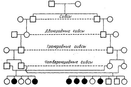
A proband is a person with whom the compilation of a pedigree begins with genealogical analysis.
Sibs is one of the children born to the same parents in relation to other children (for example, brother or sister).
2. Twin
This method consists in the study of patterns of inheritance of characters in pairs of single and twin twins. It was proposed in 1875 by Galton initially to assess the role of heredity and the environment in the development of human mental properties. Currently, this method is widely used in the study of heredity and variability in humans to determine the correlative role of heredity and the environment in the formation of various signs, both normal and pathological. It allows you to identify the hereditary nature of the trait, to determine the penetrance of the allele, to evaluate the effectiveness of the action of certain external factors on the body (drugs, training, education).
The essence of the method consists in comparing the manifestation of a trait in different groups of twins, taking into account the similarities or differences in their genotypes. Monozygous twins , developing from a single fertilized egg, are genetically identical, as they have 100% of common genes. Therefore, among monozygotic twins, there is a high percentage of concordant steam, in which the trait develops in both twins. Comparison of monozygotic twins, brought up in different conditions of the postembryonic period, allows to reveal the signs, in the formation of which the important role belongs to environmental factors. According to these signs, discordance is observed between the twins, i.e. the differences. On the contrary, the preservation of similarities between twins, despite the differences in the conditions of their existence, indicates a hereditary conditionality of the trait.
3. Population-statistical
With the help of the population-statistical method, hereditary traits are studied in large groups of the population, in one or several generations. An important point when using this method is the statistical processing of the data obtained. This method can be used to calculate the frequency of occurrence in a population of various alleles of a gene and different genotypes for these alleles, to find out the distribution of various hereditary traits, including diseases, in it. It allows studying the mutational process, the role of heredity and the environment in the formation of human phenotypic polymorphism according to normal characters, as well as in the occurrence of diseases, especially with hereditary predisposition. This method is also used to determine the significance of genetic factors in anthropogenesis, particularly in race formation.
4. Dermatoglyphic
In 1892 F.Galton as one of the methods for the study of man was proposed a method of studying the scallop skin patterns of the fingers and palms, as well as flexion palmar furrows. He found that these patterns are an individual characteristic of a person and do not change throughout life. Currently, hereditary conditionality of skin patterns is established, although the nature of inheritance is not fully understood. It is likely that the trait is inherited according to a polygenic type. Dermatoglyphic studies are important in identifying twins. The study of people with chromosomal diseases revealed specific changes in them, not only of the patterns of fingers and palms, but also of the nature of the main bending grooves on the skin of the palms. Dermatoglyphic changes in gene diseases are less studied. Basically, these methods of human genetics are used to establish paternity.
Examination of the prints of the skin pattern of the palms and feet. With the existing individual differences in the fingerprints due to the peculiarities of the development of the individual, several main classes of them are distinguished. Peculiar changes of fingerprints and palm patterns were observed in a number of hereditary degenerative diseases of the nervous system. Typical of Down's disease is a monkey (four-fingered) fold, representing a line running across the palm in the transverse direction. Currently, the method is mainly used in forensic medicine.
5. Biochemical
Hereditary diseases that are caused by gene mutations that change the structure or rate of protein synthesis, are usually accompanied by a violation of carbohydrate, protein, lipid and other types of metabolism. Hereditary metabolic defects can be diagnosed by determining the structure of the altered protein or its quantity, identifying defective enzymes or detecting the intermediate metabolic products in the extracellular body fluids (blood, urine, sweat, etc.). For example, the analysis of amino acid sequences of mutationally altered protein chains of hemoglobin revealed several hereditary defects underlying a number of diseases? hemoglobinosis. Thus, in sickle cell anemia in humans, abnormal hemoglobin due to mutation differs from the normal one by replacing only one amino acid (glutamic acid with valine).
In the practice of health care, in addition to identifying homozygous carriers of mutant genes, there are methods for identifying heterozygous carriers of certain recessive genes, which is especially important for medico-genetic counseling. Thus, phenotypically normal heterozygotes for phenylketonuria (a recessive mutant gene; amino acid metabolism of phenylalanine is disturbed in homozygotes, which leads to mental retardation) after ingestion of phenylalanine, its elevated blood levels are detected. In hemophilia, heterozygous carriage of the mutant gene can be established by determining the activity of the enzyme that has been altered as a result of the mutation.
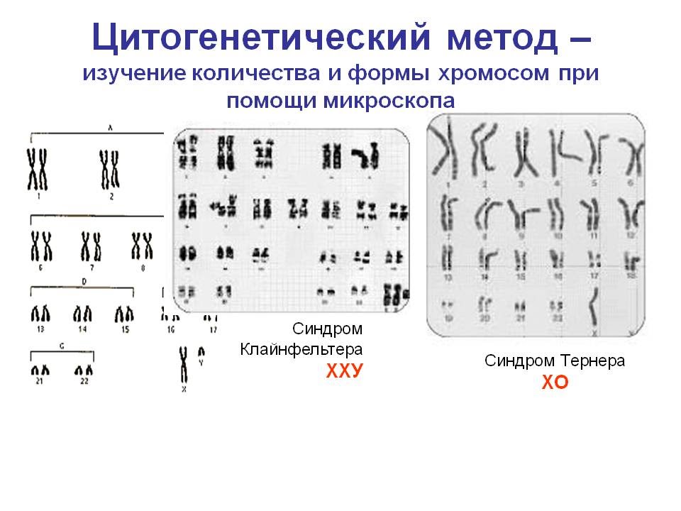
6. Cytogenetic
The cytogenetic method is used to study the normal human karyotype, as well as in the diagnosis of hereditary diseases associated with genomic and chromosomal mutations. In addition, this method is used in the study of the mutagenic action of various chemicals, pesticides, insecticides, drugs, etc.
During the period of cell division into metaphase stages, chromosomes have a clearer structure and are available for study. The diploid set of a person consists of 46 chromosomes: 22 pairs of autosomes and one pair of sex chromosomes (XX? In women, XY? In men). Usually, human peripheral blood leukocytes are examined, which are placed in a special nutrient medium, where they divide. Then prepare the preparations and analyze the number and structure of the chromosomes. The development of special coloring methods significantly simplified the recognition of all human chromosomes, and together with the genealogical method and the methods of cell and genetic engineering, made it possible to correlate genes with specific chromosome regions. The integrated application of these methods underlies the mapping of human chromosomes. Cytological monitoring is necessary for the diagnosis of chromosomal diseases associated with ansuploidy and chromosomal mutations. The most common Down syndrome (trisomy 21 st chromosome), Klinefelter syndrome (47 XXY), Shershevsky syndrome? Turner (45 XO) and others. Loss of a portion of one of the homologous chromosomes of the 21st pair leads to blood disease? chronic myeloid leukemia.
Cytological studies of the interphase nuclei of somatic cells can reveal the so-called Barry body, or sex chromatin. It turned out that sex chromatin is normal in women and absent in men. It is the result of heterochromatization of one of the two X chromosomes in women. Knowing this feature, you can identify gender and identify an abnormal number of X chromosomes.
Detection of many hereditary diseases is possible even before the birth of a child. The method of prenatal diagnosis is to obtain amniotic fluid, where the cells of the fetus are located, and in the subsequent biochemical and cytological determination of possible hereditary anomalies. This allows you to make a diagnosis in early pregnancy and make a decision about her continuation or termination.
With the help of these methods, the heredity and variability of somatic cells are studied, which compensates for the impossibility of applying a hybridological analysis to a person. These methods, based on the reproduction of these cells in artificial conditions, analyze the genetic processes in individual cells of the body, and thanks to the usefulness of the genetic material, use them to study the genetic patterns of the whole organism.
Hybrid cells containing 2 complete genomes usually divide chromosomes, preferably one of the species, upon division. Thus, cells with the desired set of chromosomes can be obtained, which makes it possible to study the linkage of genes and their localization in certain chromosomes.
Thanks to the methods of somatic cell genetics, it is possible to study the mechanisms of the primary action and interaction of genes, the regulation of gene activity. The development of these methods determined the possibility of accurate diagnosis of hereditary diseases in the prenatal period.
8. Simulation method
Studying human diseases in animals that can suffer from these diseases. It is based on the Vavilov law on homologous series of hereditary variability, for example, hemophilia linked to sex, can be studied in dogs, epilepsy in rabbits, diabetes, muscular dystrophy in rats, and lip and palate cleft in mice
Models in biology are used to model biological structures, functions, and processes at different levels of the organization of life: molecular, subcellular, cellular, organ-systemic, organismic, and population-biocenotic. It is also possible to model various biological phenomena, as well as the living conditions of individual individuals, populations and ecosystems.
In biology, three types of models are mainly used: biological, physico-chemical, and mathematical (logical-mathematical). Biological models reproduce certain conditions or diseases found in humans or animals in laboratory animals. This makes it possible to study experimentally the mechanisms of the occurrence of a given condition or disease, its course and outcome, to influence its course. Examples of such models are artificially induced genetic disorders, infectious processes, intoxication, reproduction of the hypertensive and hypoxic states, malignant neoplasms, hyperfunction or hypofunction of certain organs, as well as neuroses and emotional states. To create a biological model, various methods of influence on the genetic apparatus, infection with microbes, the introduction of toxins, the removal of individual organs or the introduction of their metabolic products (for example, hormones), various effects on the central and peripheral nervous systems, exclusion of certain substances from food, in artificially created habitat and many other ways. Biological models are widely used in genetics, physiology, and pharmacology.
9. Immunogenetic
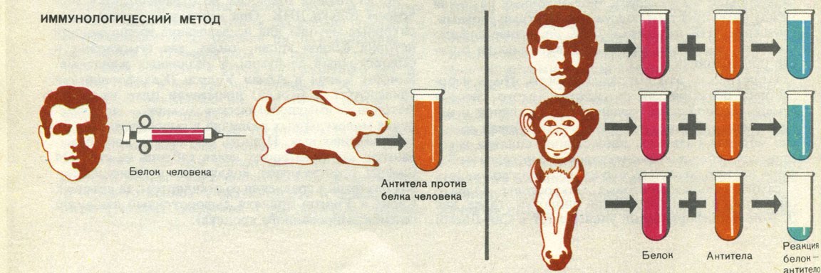
Immunological (serological) method includes the study of serum, as well as other biological substrates for the detection of antibodies and antigens.
There are serological reactions and immunological methods using physical and chemical labels. Serological reactions are based on the interaction of antibodies with antigens and the registration of the phenomena accompanying it (agglutination, precipitation, lysis). In immunological methods, physical and chemical labels are used that are included in the antigen-antibody complex being formed, allowing the formation of this complex to be recorded.
Classical serodiagnosis is based on the detection of antibodies to the identified or suspected pathogen. A positive result of the reaction indicates the presence in the test serum of antibodies to the antigens of the pathogen, a negative result indicates the absence of such.
Serological reactions are semi-quantitative and allow to determine the antibody titer, i.e. the maximum dilution of the test serum, which still has a positive result.
Detection of antibodies to the causative agent of a number of infectious diseases in the test serum is not sufficient for diagnosis, since it may reflect the presence of post-infectious or post-vaccination immunity. That is why they study paired serum - taken in the first days of the disease and after 7-10 days. In this case, an increase in antibody titer is assessed. Diagnostic significant increase in antibody titer in the test serum relative to the initial level is 4 times or more. This phenomenon is called seroconversion.
In exotic infectious diseases, as well as in hepatitis, HIV infection and some other diseases, the very fact that antibodies are detected indicates that the patient is infected and has diagnostic value.
Human genetics studies the phenomena of heredity and variability in human populations, the features of inheritance of characters in normal conditions and their changes under the influence of environmental conditions.
Man as an object of genetic analysis. The study of human genetics is associated with great difficulties:
One of the first conditions for hybridological analysis in humans is impracticable, since experimental marriages in humans are impossible. People marry without pursuing any "experimental" goals.
23 pairs of chromosomes complicates genetic and cytological mapping, which in turn reduces the possibilities of genetic analysis.
To change one generation takes an average of 30 years. Consequently, a geneticist cannot observe more than one or two generations.
The family size is currently so small that it does not allow the analysis of the splitting of characters in the offspring within the same family.
For humans, the concept of “environment” is broader than for animals and plants. In addition to such factors as exercise, nutrition, living conditions, climate, the environment of a person is his social life, and it is not amenable to change at the request of genetics.
Genealogy in the broadest sense of the word pedigree - the genealogical method - the pedigree method. It was introduced at the end of the 19th century by F. Galton and is based on the construction of pedigrees and tracking of the disease (or trait) in a family or genus, indicating the type of kinship between the members of the pedigree. Currently, it is the most universal and widely used in solving theoretical and applied problems.
1) is this trait hereditary
2) the type of inheritance and gene penetrance
3) to assume the genotype of persons of the pedigree
4) determine the probability of having a child with the disease being studied
5) intensity of the mutation process
6) used to compile genetic maps of chromosomes
Thus, the goal of the genealogical method comes down to clarifying kinship and tracking a trait or disease among close and distant, direct and indirect relatives. Technically, it consists of the following steps.
Stages of genealogical analysis:
1) collection of data on all relatives of the subject (history)
2) building a pedigree
3) pedigree analysis and conclusions
The difficulty of collecting anamnesis is that the proband should know most of its relatives and their state of health. A proband is a person who has applied to a medical genetic consultation in respect of which a pedigree is being built, and from whom information is obtained regarding the same disease from relatives. Sibs - brothers and sisters of the proband.
1. Autosomal dominant
1. patients in each generation
2. sick child with sick parents
3. equally sick men and women
4. inheritance goes vertically and horizontally
5. The probability of inheritance 100%, 75% and 50%.
These signs will appear only with complete domination, so polydactyly, freckles, curly hair, brown eye color, etc. are inherited in humans. In case of incomplete dominance, an intermediate form of inheritance will appear. With incomplete gene penetrance, patients may not be in every generation.
2. Autosomal recessive
Most often, the probability of inheritance of a disease of this type is 25%, since, due to the severity of the disease, patients either do not live up to childbearing age, or do not marry. So inherited phenylketonuria, sickle cell anemia, blue eyes, etc.
Examples: hemophilia, color blindness, hereditary anemia, muscular dystrophy, etc.
4. X-coupled dominantthe type of inheritance is similar to autosomal dominant, except that a man passes this trait to all daughters
Example: rickets resistant to vitamin D treatment, tooth enamel hypoplasia, follicular hyperkeratosis.
Examples: auricle hypertrichosis, membranes between the second and third toes on the legs; gene that determines the development of the testes. Hollandic signs are not significant in human hereditary pathology.
II. Cytogenetic method
Currently, the cytogenetic method in genetics occupies a significant place. The use of this method allows to study the morphological structure of individual chromosomes and the karyotype as a whole, to determine the genetic sex of the body, as well as to diagnose various chromosomal diseases associated with a violation of the number of chromosomes or a violation of their structure. The method is used to study the mutation process and the compilation of genetic maps of chromosomes. The most common method is used in the prenatal diagnosis of chromosomal diseases.
The cytogenetic method is based on the microscopic study of the karyotype and includes the following steps:
Cultivation of human cells (often lymphocytes) on artificial nutrient media
Stimulation of mitosis by phytohemagglutinin (PHA)
Adding colchicine (destroys the threads of the spindle division) to stop mitosis at the metaphase stage
Treatment of cells with a hypotonic solution, as a result of which chromosomes are scattered and lie loose
Chromosome staining
Microscopic examination (computer programs).
Cytological maps of chromosomes -
Chromosome Genetic Maps, ie schemes describing the order of the location of genes and other genetic elements in the chromosome, indicating the distance between them. The genetic distance is determined by the frequency of recombination between homologous chromosomes (the distance between the genes is directly proportional to the frequency of crossing-over) and is expressed in centimeters (cM). One centimorganis corresponds to a recombination frequency equal to 1% .............. Such genetic maps, in addition to gene inventory, answer the question about the involvement of genes in the formation of individual traits of an organism.
The method allows to detect genomic (for example, Down's disease) and chromosomal (feline cry syndrome) mutations. Chromosomal aberrations are designated by the number of a chromosome, a short or long arm, and an excess (+) or shortage (-) of genetic material.
The method consists in studying the patterns of inheritance of characters in pairs of monozygous and dizygotic twins. It allows to determine the correlative role of heredity (genotype) and the environment in the manifestation of various signs, both normal and pathological. Allows you to identify the hereditary nature of the trait, determine the penetrance of the allele, assess the effectiveness of the action of certain external factors on the body (drugs, training, education).
The method consists in comparing the manifestation of a trait in different groups of twins, taking into account the similarities or differences in their genotypes.
There are mono - and dizygotic twins.
Monozygous twins develop from a single fertilized egg. They have exactly the same genotype, because have 100% of common genes. And if they differ in phenotype, then this is due to the influence of environmental factors.
Dizygotic twins develop after fertilization of several simultaneously mature eggs by sperm. Gemini will have a different genotype and their phenotypic differences will be due to both the genotype and environmental factors.
The percentage of similarity of a group of twins on the studied trait is called concordance, and the percentage of difference is called discordance. Since monozygous twins have the same genotype, the trait develops in both twins, their concordance is higher than that of dizygotic ones. Comparison of monozygous twins, brought up in different conditions, allows to reveal the signs, in the formation of which the essential role belongs to environmental factors, according to these signs discordance is observed between twins, i.e. the differences.
To assess whether heredity and environment in the development of a particular trait, use the Holzinger formula:
With MZ - With DZ
H = --------------------- x 100 E = 100 - H
H - the role of heredity, E - the role of the environment
As the theoretical foundations of the twin method were developed, a special section of these studies was gradually formed - the method of control by partner. Allows you to evaluate the therapeutic effect of new pharmacological agents for different routes of administration, explore the phases of their action, show the differences in pharmacokinetics of new and old drugs). The method is used for susceptibility to various diseases: coronary artery disease, peptic ulcer, rheumatism, infectious diseases, tumors.
IV. Population-statistical method
With its help, hereditary traits are studied in large populations, in one or several generations. It allows you to determine the frequency of occurrence in a population of different alleles of a gene and different genotypes for these alleles, to determine the distribution of various hereditary traits, including diseases. It allows you to study the mutation process, the role of heredity and the environment in the occurrence of diseases, especially with hereditary predisposition. The essential point of using this method is the statistical processing of the data obtained on the basis of the Hardy-Weinberg law of genetic equilibrium.
The mathematical expression of the law is the formula (pA + qa) 2 where p and q are the frequencies of occurrence of alleles A and a of the corresponding gene. The disclosure of this formula makes it possible to calculate the frequency of occurrence of people with different genotypes, and first of all heterozygotes - carriers of the hidden recessive allele: p 2 AA + 2pq + q 2 aa.
However, before talking about the practical application of these formulas, we should note the conditions for the emergence of an equilibrium of genotypes in populations:
1) Presence of panmixia, i.e. random selection of married couples
2) Lack of allele influx caused by mutation pressure
3) Absence of outflow of alleles caused by selection.
4) Equal fertility of heterozygotes and homozygotes
5) Generations should not overlap in time.
6) The population size should be large enough.
Well-known genetics point out that although this set of conditions cannot be met in any particular population, in most cases the calculations according to the Hardy-Weinberg law are so close to reality that this law turns out to be quite suitable for analyzing the genetic structure of populations.
Example……..
For example, HbS homozygous in Belarus practically does not occur, and in West African countries their frequency varies from 25% in Cameroon to 40% in Tanzania. The study of the distribution of genes among the population of different geographic zones (genogeography) makes it possible to establish the centers of origin of various ethnic groups and their migration, to determine the degree of risk of hereditary diseases in certain individuals.
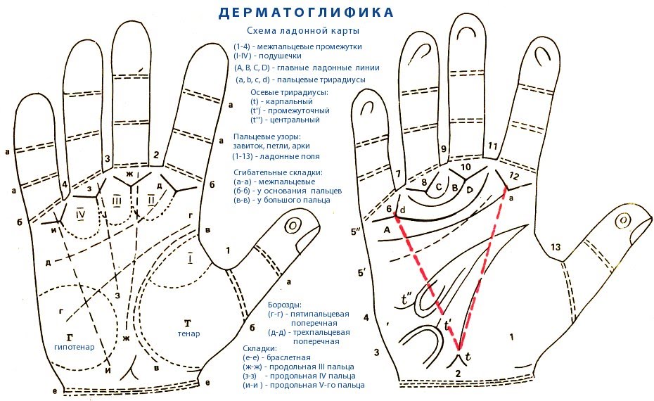
V. Method of dermatoglyphics and palmoscopy (fingerprinting)
In 1892, Galton was proposed as one of the methods for studying human genetics - This is the method of studying the skin scallop patterns of the fingers and palms, as well as flexor palmar furrows. These patterns are an individual characteristic of a person and do not change during his life; they are restored after injuries (burns).
Example (Galton, Gioconda)
It has now been established that the trait is inherited according to a polygenic type and the mother has a great influence on the character of the finger and palm patterns through the mechanism of cytoplasmic heredity.
The method is widely used in forensic science, identification of the zygosity of twins, the establishment of paternity. Characteristic changes in these patterns are observed in some chromosomal diseases (sm Down, Klinefelter, Sher.-Turner).
VI. Biochemical methods
Allows you to study hereditary diseases caused by gene mutations - the causes of metabolic diseases (phenylketonuria, sickle cell anemia). Using this method, more than 1000 congenital metabolic diseases have been described, and for many of them a defect in the primary gene product has been identified. The most common among these diseases are diseases associated with deficiency of enzymes, structural, transport or other proteins.
The method is based on studying the activity of enzyme systems: either by the activity of the enzyme itself, or by the number of final reaction products catalyzed by this enzyme.
Defects of enzymes are determined by determining the content in the blood and urine of metabolic products that are the result of the functioning of this protein. The lack of the final product, accompanied by the accumulation of intermediate and by-products of disturbed metabolism, indicates a defect in the enzyme or its deficiency in the body.
Using biochemical stress tests, heterozygous carriers of pathological genes, such as phenylketonuria, can be identified. The person being tested is administered intravenously a certain amount of the amino acid phenylalanine and at regular intervals determine its concentration in the blood. If a person is homozygous for the dominant gene (AA), then the concentration of phenylalanine in the blood returns to the control level rather quickly, and if he is heterozygous (Aa), then the decrease in the concentration of phenylalanine goes twice as slowly.
Similarly, tests are conducted that identify susceptibility to diabetes, hypertension and other diseases.
VII. Recombinant DNA Methods
Allow to analyze DNA fragments, to find and isolate individual genes and gene segments and to establish in them the sequence of nucleotides. This method includes the method of DNA cloning. The term “cloning” means that a gene has been cloned, it has been isolated by special techniques, its structure has been studied, and gene cloning also means that a protein is known, the synthesis of which is controlled by the corresponding gene. “Genomic libraries” and international data banks are created on the basis of cloned genes. Any specialist in the world can easily enter these data banks and use the information collected there for research purposes. These genomic libraries are widely used in the implementation of the “human genome” program. (A collection of DNA fragments from the entire genome)
Thanks to the success achieved under this program, it became possible to realistically assess the function of genes in the human body. Although information for more than a quarter of genes is not yet available, for two thirds of the genes, it is either fully established or can be roughly indicated. Also, extremely interesting information was obtained on the involvement of genes in the formation and functioning of individual organs and tissues of the human body. It turned out that the largest number of genes is necessary for the formation of the brain and maintaining its activity, and the smallest for the creation of red blood cells - only 8 genes. This information will help to understand the genetic programs of development and functioning of the human body, the causes of cancer and aging. Identification of the molecular basis of diseases will help to transfer to a new level the methods of their early diagnosis, and, therefore, to more subtly and successfully fight diseases. Methods such as, for example, targeted delivery of drugs to affected cells, the replacement of diseased genes with healthy ones, and many others become part of the arsenal of modern medicine.
Viii. Somatic cell genetics methods
With the help of these methods, the heredity and variability of somatic cells are studied, which largely compensates for the impossibility of applying the hybridological method to humans.
Human somatic cell cultures are obtained from biopsies (peripheral blood, skin, tumor tissue, embryo tissue, cells from amniotic fluid).
In human genetics, the following four methods are used.
1. Simple cultivation - cells suitable for cytogenetic, biochemical, immunological and other studies.
2. Cloning - getting the descendants of a single cell. It makes it possible to carry out a biochemical analysis of hereditary processes in genetically identical cells.
3. Selection of somatic cells using artificial media is used to select mutant cells with certain properties, selection of hybrid cells. The method is widely used to study gene mutations (mechanisms, spontaneous and induced frequency).
4. Hybridization of somatic cells is based on the fusion of co-cultured cells of different types. With the introduction of cell culture RNA-sod. Sendai virus inactivated by ultraviolet irradiation - the frequency of hybridization increases significantly. Heterokaryons -2 nuclei of different cells in the same cytoplasm. After mitosis, two mononuclear cells are formed - synkaryons - a real hybrid cell containing the chromosomes of both source cells. In the future, there is a gradual removal of the chromosomes of the organism, whose cells have a slower reproduction rate.
The loss of chromosomes is random and therefore, among a large number of hybrids, you can always find a cell that has retained any one human chromosome.
Using a suitable selective system, one can select cells with a certain enzymatic activity and localize the gene of this enzyme on a specific chromosome.
The method is used to study the problems of coupling and localization of genes.
It is possible to study the mechanisms of the primary action and interaction of genes, the regulation of gene activity. The method allows you to widely study the pathogenesis of hereditary diseases at the biochemical and cellular level.
Ix. Creating models of human hereditary diseases using transgenic
animals.
Biological modeling of hereditary diseases is a large section of experimental biology and genetics. The principle of biological modeling of gene mutations is based on the law of homologous series in hereditary variation discovered by N.I. Vavilov. In animals, there are mutations that cause the same pathological effect as in humans (mice, rabbits, dogs, hamsters, mice). Among the hereditary anomalies in animals there are such diseases as hemophilia, achondroplasia, muscular dystrophy, diabetes and many others that form the basis of the human hereditary pathology.
Methods are based on the introduction of foreign genes into germ cells.
Like any model, mutant lines of transgenic animals cannot fully reproduce the hereditary disease; therefore, certain specific fragments are modeled in order to study the primary mechanism of gene action, the pathogenesis of the disease, and the principles of its treatment.
The basic laws of heredity established for living organisms are universal and fully true for humans. However, as an object of genetic research, a person has its advantages and disadvantages.
For people it is impossible to plan artificial marriages. As early as 1923, N. K. Koltsov (1923) noted that "... we cannot experiment, we cannot force Nezhdanov to marry Chaliapin only to see what kind of children they will have." However, this difficulty is surmountable, thanks to a targeted sampling of a large number of married couples that correspond to the goals of this genetic study.
A large number of chromosomes (46) greatly complicates the possibilities of genetic analysis of man. The development of new methods of working with DNA (the method of hybridization of somatic cells and some others) eliminate this difficulty.
Due to the small number of descendants (in the second half of the 20th century, most families gave birth to 2-3 children), it is impossible to analyze the splitting in the offspring of one family. However, in large populations, it is possible to select families with traits of interest to the researcher. In addition, in some families, certain signs have been traced over many generations, and in such cases genetic analysis is possible. Another difficulty is related to the duration of a generation change in a person. One generation takes an average of 30 years, and therefore a geneticist cannot observe more than one or two generations.
For humans, genotypic and phenotypic polymorphism is characteristic. Manifestations of many symptoms and diseases largely depend on environmental conditions. It should be noted that the concept of "environment" for humans is broader than for plants and animals. Along with nutrition, climate and other abiotic and biotic factors for a person, social factors are also a medium that are difficult to change at the request of the researcher. However, a person as a genetic object is widely studied by doctors of all specialties, which often helps to establish various hereditary deviations.
So, human genetics has a number of features:
a) experimental marriages are prohibited in humans;
b) a small number of descendants are born;
c) there is a late puberty and a longer generational change (25-30 years);
d) a person has a complex karyotype (many chromosomes and linkage groups);
d) the inability to create the same living conditions of the study.
Despite these difficulties, human genetics is better studied today than the genetics of many other organisms.
Currently, interest and attention to the study of human genetics is actively increasing. Thus, the global international program "Human Genome" has as its task the study of the human genome at the molecular level. To solve it, all the latest modern methods of genetics and medicine are used.
The main methods of studying human heredity:
1. Clinical genealogical method.It was introduced at the end of the XIX century F.
Galton and is based on the compilation and analysis of pedigrees.
The genealogical method is the method of studying the pedigrees, by means of which the distribution of the disease (trait) in the family or in the gens is traced, indicating the type of kinship between the members of the pedigree.
Empirical observations on the pedigrees in which the transmission of pathological signs or diseases was noted have been known for a long time. The use of family analysis to study human pathology in the XVIII-XIX centuries. can be considered as prerequisites for the formation of the genealogical method. Its further improvement proceeded both along the line of compilation of pedigrees, and especially with respect to methods of statistical data analysis.
The genealogical method refers to the most universal methods in medical genetics. This method is often referred to as clinical genealogical, since it is a question of studying the pathological signs (diseases) in a family with the help of clinical examination techniques. It is widely used in solving theoretical and applied problems:
To establish the hereditary nature of the trait;
In determining the type of inheritance of a trait or disease;
To assess gene penetrance;
When analyzing linkage of genes and chromosome mapping;
When studying the intensity of the mutation process;
When deciphering the mechanisms of interaction of genes.
The genealogical method takes a special place in medical and genetic counseling, being sometimes decisive or the only method:
To clarify the nature of the disease;
When making a diagnosis of a hereditary disease;
For the differential diagnosis of hereditary diseases;
In assessing the prognosis of the disease;
When calculating the risk for offspring;
To select adequate and justified methods for prenatal diagnosis.
In the clinical - genealogical method, two successive stages are distinguished:
1. compilation of the pedigree and its graphic image;
2. genetic analysis of the data.
Consider in detail each of these stages.
Drawing a pedigree. The collection of information about the family begins with the proband - the individual who is the subject of interest of the doctor (researcher) to this particular pedigree. Most often it is the patient or carrier of the studied trait. However, healthy individuals can apply for genetic counseling for various reasons, and in this case it is better to use the term “counseling”. Children of the same parent couple (brothers and sisters) are called siblings. If siblings have only one common parent, they are called half siblings. There are single uterine (common mother) and half-siblings (common father) half siblings.
Usually a pedigree is collected on one or more grounds. Most often, a patient or a consultant is concerned about a particular disease or symptom.
It is necessary to strive for the most complete compilation of pedigrees in ascending, descending and lateral directions. This task is not as simple as it may seem at first glance. The more generations involved in the pedigree, so it is more extensive. This can lead to a distortion of the information received, and then to the inaccuracy of the pedigree itself and conclusions made on the basis of its analysis.
It is necessary to collect information concerning not only the presence of a particular disease or pathological sign in the family, but also information on all cases of diseases occurring among family members. It is important to obtain information on spontaneous abortions, stillbirths and early infant mortality. They may be directly related to the substance of the issue and play an important role in assessing the forecast. You should always get at least basic data from both sides (paternal and maternal), even if we are talking about an autosomal dominant disease inherited from one of the parents.
For the analysis and visual presentation of the collected information using a graphical image of the pedigree. To do this, use standard symbols (Fig. 3.3) and techniques. However, depending on the tasks, goals and characteristics of the pedigree, the compiler may use his own designations with their obligatory explanation under the figure. To clarify the principles of designation and compilation of pedigrees, we give two examples (Fig.3.4, 3.5). As can be seen from these figures, generations are denoted by Roman numerals from top to bottom. Usually they are placed to the left of the pedigree. The last generation of ancestors, for which information is collected, receives the designation I generation. Arabic numerals numbered offspring of one generation (the whole row) from left to right sequentially. Brothers and sisters are ranked in order of birth (from older to younger). Thus, each member of the pedigree has its own cipher, for example, II-3, III-5. In those cases when the spouse is not examined for the presence of the considered feature and its pedigree is not given, it is advisable not to depict it at all. All individuals of the same generation should be strictly in one row, so it is preferable to draw a family tree on lined paper. Hanging characters between generations is a blunder. If the pedigree is very extensive, then different generations are arranged not in horizontal rows, but concentric.
A graphic representation of the pedigree must be accompanied by a “pedigree legend”, which is an essential element of the description of the pedigree and includes:
1) a description of the state of health of a member of the pedigree, information about which is important for understanding the nature of the inheritance of the disease (trait) or the characteristics of its clinical manifestation;
2) the age of onset and the nature of the course of the disease in those affected;
3) the cause of death and age at the time of death of a member of the pedigree;
4) a description of the methods of diagnosis and identification of diseases, a list of sources of medical and other information.
When applying the genealogical method, it is important to note in the pedigree personally examined for the presence of a trait (this can also be equated with obtaining information from an objective source, such as a medical history) and unexplored, information about which is obtained from the responses of the proband or relatives, as well as from questionnaires.
We must strive to obtain the most complete and objective primary material, which is the basis of statistical and genetic analysis and, accordingly, a guarantee of the correctness and accuracy of the resulting conclusions.
Genealogical analysis of the pedigree.The goal of genealogical analysis is to establish genetic patterns.
The first task in the analysis of the pedigree is to establish the hereditary nature of the trait. If the same trait (or disease) occurs several times in the pedigree, you can think of its hereditary nature. However, we must first exclude the possibility of phenocopy (the disease is similar to a hereditary disease, while the cause of its development is the environmental factor). For example, if a pathogenic factor acted on a woman during all of her pregnancies, then she could have several children with the same congenital anomalies. On the other hand, the effect of the same external factors (professional, household, etc.) on several members of the pedigree can also cause them to have similar diseases. Thus, if the action of similar external factors is excluded, and for representatives of different generations it is more likely to be excluded, then one can think about the hereditary nature of the disease.
After the hereditary nature of the disease (trait) is established, it is necessary to establish the type of inheritance. For this purpose, the principles of genetic analysis and various statistical methods of processing data obtained from the pedigree are used.
It is easy to understand that in most cases the calculations of the ratio of the number of sick and healthy in one particular family can give a wrong idea of the type of inheritance. This is mainly due to the random nature of the inheritance of different alleles. In each family, the ratio of sick and healthy children may differ significantly from the theoretically expected ratios characteristic of a certain type of inheritance. However, the nature of the pedigree, features of transmission of the disease (trait) in the generations, compliance with their criteria for inheritance of one type or another allow us to make a certain conclusion about the type of inheritance of the pathology (trait) in a particular family.
Only monogenic hereditary diseases, that is, diseases whose etiological factor is mutation of a single gene, are subject to Mendelian inheritance patterns. Depending on the localization and properties of the gene, autosomal dominant and autosomal recessive inheritance types are distinguished, when the gene is located in one of 22 pairs of autosomes (non-sex chromosomes), X-linked dominant and recessive inheritance types, when the gene is located in the X chromosome, Y -linked (holandric) inheritance when the gene is located on the Y chromosome, as well as mitochondrial (maternal or cytoplasmic) inheritance, when a mutation occurs in the mitochondrial genome.
1.1 Genes in the family. Inheritance Type Criteria
Autosomal dominant inheritance. If the disease is caused by a rare autosomal dominant gene, then the absolute majority of patients in the population are born in marriages between the affected and healthy spouse. In this case, one of the parents is heterozygous for the autosomal dominant gene (Aa), and the other is homozygous for the normal allele (aa). In such a marriage, the following variants of genotypes in the offspring are possible: Aa, Aa, aa, aa.
Thus, in every 50% of cases, every future child is likely to receive both allele A (and therefore be affected) and the normal allele from a sick parent and be healthy. Thus, the ratio of the number of healthy children in the offspring to the number of affected ones is 1: 1 and does not depend on the sex of the child.
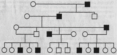
In general, the main criteria for suspecting autosomal dominant inheritance of the disease are:
the disease manifests itself in each generation without gaps (“vertical” type);
every child of a parent with an autosomal dominant disease has a 50% risk of inheriting this disease;
unaffected children of sick parents are free from the mutant gene and have healthy children;
diseases are inherited by men and women equally often and with a similar clinical picture.
In the pedigrees shown in Fig. 3.6, 3.7, one can see compliance with the criteria for autosomal dominant inheritance. Exceptions to the rule about the manifestation of autosomal dominant diseases in each generation are cases of new mutation (sporadic cases) or incomplete penetrance (manifestation) of the gene.
A sporadic is the only case of a hereditary dominant disease in the family (Fig. 3.8). It can be the result of a mutation that has re-emerged in the germ cells of one of the healthy parents. In the case of incomplete penetrance in the pedigree with an autosomal dominant type of inheritance, there will be cases of generation skipping or “skipping”, i.e., the individual will have an affected ancestor and an affected descendant, while he will be healthy (Fig. 3.9).
To date, described about 3,000 autosomal dominant traits of man.
The most commonly encountered in clinical practice following monogenic disease with autosomal dominant inheritance: familial hypercholesterolemia, Marfan syndrome, neurofibromatosis type 1 (Reklingha-uzena disease), Ehlers-Danlos syndrome, myotonic dystrophy, achondroplasia, osteogenesis imperfecta, and others.
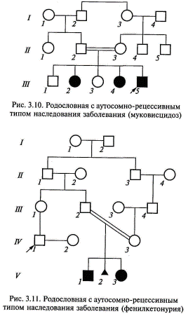
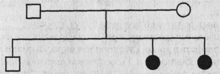
Autosomal recessive inheritance. Diseases with this type of inheritance occur only in homozygotes, which have received one recessive gene from each of the parents. It is found in the proband's sibs, but sometimes it is also found in the lateral branches of the pedigree. Characteristic of autosomal recessive diseases is an Aa x Aa marriage (both parents are healthy, but are carriers of the mutant gene). The probability of having a sick child in such a marriage is 25%. Children with recessive diseases have, as a rule, phenotypically healthy parents, and such families can be determined only after the birth of a sick child.
In the population, the meeting of two carriers of a rare autosomal recessive gene is an infrequent event, but its probability increases significantly in the case of consanguineous spouses. That is why recessive diseases often manifest themselves in kinship marriages.
The following signs are characteristic of rare autosomal recessive diseases:
parents of a sick child are usually healthy and are heterozygous carriers of the pathological allele;
boys and girls get sick equally often;
repeated risk of having a child with an autosomal recessive disease is 25%;
there is a “horizontal” distribution of patients, that is, patients are more often found within the offspring of one parental pair;
there is an increase in the frequency of sick children in kinship marriages, and, the less often autosomal recessive diseases are, the more often patients come from hereditary marriages;
married two affected parents, all children will be sick.
Examples of pedigrees with autosomal recessive inheritance are shown in Fig. 3.10, 3.11.
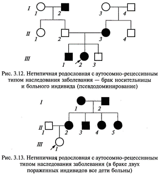
In the marriage of an affected individual and a heterozygous carrier of the same mutant allele, the risk for future children will be 50%, as with an autosomal dominant mode of inheritance, rather than 25%, as it should be with typical autosomal recessive inheritance. Therefore, this variant of autosomal recessive inheritance is called pseudo-dominance (Fig. 3.12).
The pedigree, where all the children are sick in the marriage of two affected parents, is shown in Fig. 3.13.
To date, more than 1,600 autosomal recessive diseases are known. The autosomal recessive type inherits the vast majority of hereditary metabolic diseases (fermentopathy). The most common and clinically significant diseases with an autosomal recessive mode of inheritance are: cystic fibrosis (cystic fibrosis of the pancreas), phenylketonuria, adrenogenital syndrome, many forms of hearing or vision impairment, mucopolysaccharidosis, glycogenosis.
Inheritance linked to the X chromosome. Genes localized in the sex chromosomes are differently distributed in men and women. In clinical genetics, X-linked diseases are of practical importance, i.e., such when the pathological gene is located on the X chromosome.
The distribution of the X-linked trait depends on the distribution of the X chromosome carrying the abnormal gene. Given that women have two X chromosomes, and men have one, a woman, having inherited a pathological allele, will be heterozygous, and a man will be hemizygous. This determines the main differences in the frequency and severity of damage to different sexes with X-linked inheritance, both dominant and recessive.
Dominant X-linked inheritance. Diseases with X-linked dominant inheritance are 2 times more common in women than in men. The main characteristic of X-linked dominant inheritance is that sick men pass the abnormal gene (or disease) to all their daughters and do not pass it on to their sons. A female patient transmits an X-linked dominant gene to half of her children, regardless of gender.
The main features of the X-linked dominant type of inheritance are as follows (Fig. 3.14):
the disease occurs in men and women, but in women about two times more often;
a sick man transmits a mutant allele to all his daughters and does not transmit to his sons, since the latter receive from the father the Y chromosome;
sick women transmit the mutant allele to 50% of their children, regardless of gender;
women in the case of the disease suffer less severely (they are heterozygotes) than men (which are hemizygotes).
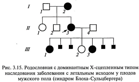
The extreme degree of unevenness of the lesion of the sexes in this type of inheritance is the existence of diseases that affect only female individuals, while male individuals who inherit the pathological allele die even in utero. Such rare diseases are referred to as X-linked dominant diseases with a lethal effect on male fetuses (for example, pigment incontinence syndrome, focal skin hypoplasia) (Fig. 3.15).
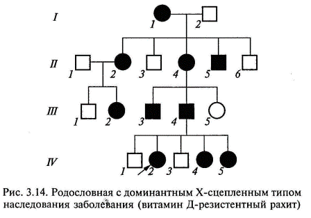
Diseases characterized by X-linked dominant inheritance include vitamin D-resistant rickets (rickets, which is not amenable to treatment with usual doses of vitamin D), rotor-spinal syndrome and other diseases.
Recessive X-linked inheritance. X-linked recessive disease (or symptom) is always manifested in men who have the corresponding gene, and in women only in cases of a homozygous state (which is extremely rare).
The main features of X-linked recessive inheritance are as follows (Fig. 3.16, 3.17):
the disease occurs mainly in males;
a symptom (disease) is transmitted from a sick father through his phenotypically healthy daughters to half of his grandchildren;
the disease is never transmitted from father to son;
subclinical signs of pathology are sometimes detected in women carriers;
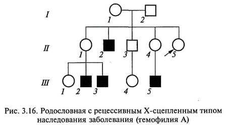
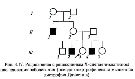
50% of the daughters will be sick, 50% of the daughters will be carriers; 50% of sons will also be sick, and 50% of sons will be healthy (for diseases that do not reduce the reproductive ability of sick men).
Severe clinical symptoms of X-linked recessive diseases in women can also be observed if they combine this monogenic defect with a chromosomal pathology, in particular, with Shereshevsky-Turner syndrome, when a woman lacks one of the two X chromosomes, as well as during accidental inactivation X chromosomes carrying the normal allele in most cells of the body.
Y-linked, or holandric, inheritance. For a long time it was believed that the Y chromosome contains only genetically inactive regions. Currently, the Y chromosome has localized about 20 genes, including the genes determining the development of the testes, responsible for spermatogenesis, controlling the growth rate, determining hair growth of the auricle, middle phalanges of the hands and some others. A trait whose genes are located on the Y chromosome is passed from the father to all boys and only boys. Pathological mutations causing impaired formation of the testes or spermatogenesis are not inherited due to the sterility of their carriers. An example of a pedigree with a Y-linked type of inheritance (ear hairiness) is presented in Fig. 3.18. „- Mitochondrial or cytoplasmic heredity. The mitochondrial genome is presented as a circular DNA molecule containing 16,569 thousand base pairs. To date, there are a number of mitochondrial DNA mutations that cause various diseases.
Since mitochondria are inherited by a child from the mother with the cytoplasm of oocytes (in mature sperm cells only
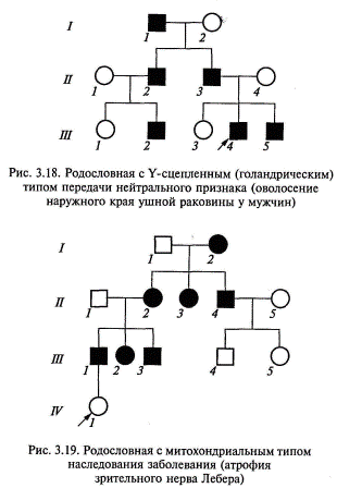
4 mitochondria), then the mitochondrial type of inheritance will be characterized by two main features (Fig. 3.19):
the disease is transmitted only from the mother to all children, regardless of the child’s gender;
sick fathers do not pass on the disease to either their sons or their daughters — all children will be healthy and the transmission of the disease will stop.
According to this type, Leber's optic nerve atrophy, mitochondrial mioencephalopathy, Kearns-Sayre syndrome and some other diseases are inherited. Currently, about 30 different diseases are described, in which mitochondrial DNA mutations are detected. Mitochondrial genes encode various respiratory chain enzymes in cells. Thus, they take part in energy processes, where they perform fairly versatile functions and are expressed in many tissues, especially in the nervous and muscular system, which explains the variety of clinical manifestations of hereditary mitochondrial diseases: myopathy, convulsions, ophthalmoplegia, and heart rhythm disturbances, as well as damage to the liver and kidneys.
Test questions and tasks
1. What questions can be solved with the help of the clinical - genealogical method?
2. List the stages of the clinical genealogical method.
3. What do the terms "proband", "sibs", "related marriage" mean?
4. List the criteria for autosomal dominant inheritance and provide examples of diseases.
5. What is a sporadic case?
6. List the criteria for an autosomal recessive inheritance type and name the diseases inherited by this type.
7. What is meant by pseudo-dominance?
8. Describe the differences between X-linked dominant and X-linked recessive types of inheritance?
9. What is the characteristic of mitochondrial inheritance?
10. What are the criteria for holandric type of inheritance?
11. List the basic principles of the genealogy, the types of genealogical schemes, symbols.
12. List the features: a) autosomal dominant inheritance; b) autosomal recessive inheritance; c) sex-linked inheritance.
13. Determine the nature of the inheritance (dominant or recessive) of an autosomal trait, write down the genotypes of all members of the pedigree below.
14. It is known that gout is inherited in an autosomal dominant manner. According to some information, the penetrance of this gene in men is 20%, and in women it is equal to 0. What is the incidence of gout in a family where both parents are heterozygous? What is the probability of gout disease in children in the case where one of the parents is heterozygous and the other is homozygous for the analyzed trait?
15. Determine the type of inheritance of an autosomal trait (dominant or recessive), indicate the genotypes of all members of the pedigree:
16. A man suffering from color blindness and deafness, married a woman, normal in sight and good hearing. They were born - a deaf daltonic son and a daltonic daughter with normal hearing. Determine the probability of having a daughter in this family with both anomalies, if it is known that color blindness and deafness are transmitted as recessive traits, but color blindness is linked to the X chromosome, and deafness is an autosomal symptom.
17. Hypertrichosis (excessive pilosis) is transmitted through the Y chromosome, and polydactyly (six-shaft) is the dominant autosomal trait. In a family where the father had hypertrichosis, and the mother had polydactyly, a daughter was born, normal for both symptoms. What is the likelihood that the next child in this family will also be without both anomalies?
18. A right-handed woman with brown eyes and normal blood married a blue-eyed right-hander male with hemophilia. They had a blue-eyed left-handed daughter with hemophilia. What is the probability that the next child in this family will be left-handed, suffering from hemophilia? It is known that brown eye color and right handedness are dominant signs, and hemophilia is a recessive X-linked trait. What eye color is possible in sick children?
19. Arachnodactyly (spider hand) is inherited as a dominant autosomal trait with a penetrance of 30%. Left-handedness - a recessive autosomal symptom with complete penetrance. Determine the probability of having a child with two anomalies in the family, in which both parents are heterozygous for both pairs of traits. 10. Analyze and determine the nature of the inheritance of the trait according to the pedigree below:
The symptom is linked to the floor. In which of the sex chromosomes is the gene located? Indicate its dominance or recession.
20. The proband suffers from night blindness. His two brothers are sick too. In line with his father, a proband was not suffering from night blindness. Mother probanda sick. Her two sisters and two brothers are healthy; their children are also healthy. Maternal grandmother was ill, grandfather is healthy; grandmother's sister is ill, and brother is healthy. Great-grandfather (grandmother's father), his sister and brother, as well as great-grandfather and his brother, who had a sick daughter and two sick sons, were also sick. The proband's wife, her parents and relatives are healthy. Determine the probability of birth of sick children in the family of the proband
21. Spouses with normal vision have four children: two daughters and two sons. The first daughter has normal vision; she has three sons, two of whom are color blind. The second daughter and her five sons have normal vision. The first son suffers from color blindness, and his two daughters and two sons see normally. The second son and his four sons have normal vision. What are the genotypes of grandparents, their children and grandchildren? Make a family tree of this family.
22. Penetration of schizophrenia in heterozygotes is 20%, in homozygotes - 100%. A man suffering from periodic exacerbations of schizophrenia marries a healthy woman. It is known that relatives of the wife had no such pathology. Her husband's grandmother was ill, but his parents are healthy. What is the forecast for this family? 14. A healthy girl aged 22, one of whose parents has diabetes, turns to genetic counseling for a prediction about herself and her future children. Make this prediction. Maximum penetrance of diabetes mellitus (20%) is achieved with an increase in life expectancy. Consider the sign as dominant.
2 Cytogenetic method. The main indications for cytogenetic studies are:
1) prenatal diagnosis of the sex of the fetus in families, burdened with diseases linked to the X chromosome;
2) undifferentiated oligophrenia (dementia);
3) habitual miscarriages and stillbirths;
4) multiple congenital malformations in a child;
5) infertility in men;
6) violation of the menstrual cycle (primary amenorrhea);
7) prenatal diagnosis at the age of mother over 35 years. Materials for pathogenetic research can be: peripheral blood cells (lymphocytes); skin fibroblasts; cells obtained by amniocentesis or chorionic biopsy; cells of abortuses, stillborn, etc.
The basis of the method is a microscopic study of human chromosomes. Cytogenetic studies have become widely used since the beginning of the 20s. XX century. to study the morphology and counting of human chromosomes, the cultivation of leukocytes to obtain metaphase plates.
The development of modern human cytogenetics is associated with the names of cytologists D. Tio and A. Levan. In 1956, they were the first to establish that a person has 46, and not 48, as was previously thought, chromosomes. This event marked the beginning of a broad study of human mitotic and meiotic chromosomes.
In 1959, the French scientists D. Lejeune, R. Turpen and M. Gautier established the chromosomal nature of Down's disease. In the years that followed, many of the common chromosomal diseases in humans were described. Cytogenetics has become the most important section of practical medicine. Currently qi
this genetic method is used to diagnose chromosomal diseases, compile genetic maps of chromosomes, study the mutation process, etc.
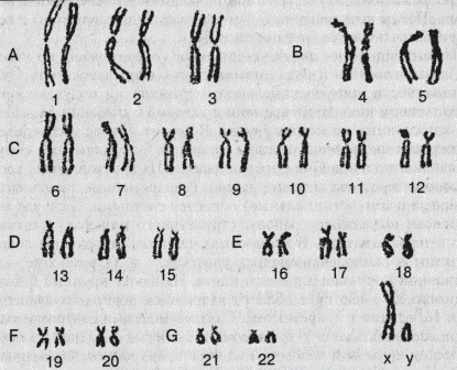
In 1960, the first International Classification of Human Chromosomes was developed in Denver (USA). It was based on the size of the chromosomes and the position of the primary waist - the centromere. All chromosomes are divided into metacentric, submetacentric and acrocentric in form and divided into 7 groups, designated by Latin letters A, B, C, B, E, F and G. Each pair of chromosome is designated by a sequence number from 1 to 23, sex chromosomes are separately distinguished X and Y (Fig. 3) In women, there are two X chromosomes, in men, the X and Y chromosomes. X-chromosome in women does not differ from the autosomes of group C; The acrocentric Y chromosome, similar to group C chromosomes, has no satellites. The length of the short arm can vary considerably. The autosomes of groups C and B contain in their short arms regions of the nucleolar organizer.
In 1971, at the 1st Prague Conference of Geneticists, in addition to the Denver classification, methods for the differential staining of chromosomes were presented, thanks to which each chromosome acquires its own unique pattern, which helps in accurate identification.
Basic information about the morphology of human chromosomes was obtained by studying them in mitosis metaphases and meiosis prophase metaphase. It is important that the number of dividing cells was high enough. The most important cytogenetic work was performed on peripheral blood lymphocytes, since the cultivation of lymphocytes for 2-3 days in the presence of phytohemagglutinin allows us to obtain many metaphase plates for chromosomal analysis.
Single-layer metaphase plates with separate chromosomes are subjected to cytogenetic analysis. For this, dividing cells are treated with colchicine and some other chemical substances (hypotonic saline solution, methanol-acetic acid fixer, etc.).
An important step in cytogenetic analysis is the color of the preparations obtained. It is carried out by simple, differential and fluorescent methods.
Simple coloring provides group identification of chromosomes. It is used to quantitatively account for chromosomal abnormalities in determining the mutagenicity of the medium (the effects of radiation, chemical mutagens, etc.). With this type of staining, many chromosomal diseases were discovered, as well as chromosomal aberrations, causing spontaneous abortions, congenital malformations, carcinogenesis, etc.
In the 70s. XX century. in medical practice, methods of differential staining began to be used, revealing the structural heterogeneity of chromosomes in length, which is expressed in the form of alternation of light and dark bands (eu- and heterochromatic regions). It is noted that the length and pattern of the bands are specific for each chromosome.
Differential staining of chromosomes can be carried out in a number of ways. Initially, acriquiniptifluorescent alkylating agent (C-method) was used.
Its action is based on the ability of metaphase chromosomes to differentially connect fluorochromes. After staining with Akrikhin, the segments acquire a bright fluorescent glow. The pattern of each chromosome is specific in number, size, and position of different fluorescent segments, which ensures the identification of all chromosomes. Using this staining method, chromatin with a higher content of at-pairs can be identified, since they are more fluorescent. A specific advantage of the Q - method is that it allows even in the interphase nucleus to identify the Y chromosome by a bright glow. To view these drugs using a fluorescent microscope.
Later, a method for coloring chromosomes without fluorescent dyes - G-coloring (Giemsa dye) was developed. After preincubation in saline, the chromosomes are treated with protease. As a result, the chromosomes acquire a segmented appearance due to the alternation of dark and light colored areas. The mechanism for the formation of segments is not yet clear. It is assumed that the colored segments are heterochromatic regions with repetitive DNA sequences, and the unstained segments are euchromatic regions with coding DNA sequences.
The varieties of differential staining according to the Gyms method are R-dyeing and C-dyeing. These varieties of differential staining are obtained with a certain change in the time and conditions of incubation of preparations stained by the Giemsa method. In the first case, the distribution of stained and unstained segments will be the opposite of what is observed with G- and Q-staining. On R-stained chromosomes, heterochromatic regions (centromere, centromeric and interstitial) remain bright. In the case of C-staining, areas of structural or optional heterochromatin are identified. In human chromosomes, these areas are located in the near-centromeric sites, and in the Y-chromosome in the distal half of the long arm. The largest C-chromatin blocks are found in the area of the secondary constrictions of the autosomes 1.9 and 16, as well as in the Y-chromosome. The Y chromosome and autosome 2 have the smallest centromeric blocks. One of the features of human chromosomes is asynchrony (non-simultaneity) replication in length. Each chromosome has early and late replicating sites. To identify the replication sequence is used 5-bromodeoxyuridine - thymine analogue. The parts that included it are stained weakly. 5-bromo-deoxyuridine is also used for differential staining of sister chromatids, if it is administered for the full cell cycle. In this case, the newly formed chromatid will turn on this thymine analogue and will be slightly colored, while the other (old) will stain intensively. This method allows you to identify areas of sister chromatid exchanges (SCO).
When exposed to various mutagenic factors, the number of CXOs increases, therefore, this method is beneficial for studying the mutation process in humans.
Advances in human molecular cytogenetics allow us to develop new methods for studying chromosomes. So, it should be noted the method of fluorescent in situ hybridization (FISH-method), which gives FISH the opportunity to explore a wide range of issues from the localization of the gene to the decoding of complex rearrangements between several chromosomes. The method can also be used to diagnose aneuploidy in interphase nuclei.
Thus, the combination of cytogenetic and molecular genetic methods in human genetics makes the possibilities of diagnosing chromosomal abnormalities almost unlimited.
Questions and tasks
1. Define the following terms: chromosome, chromatid, chromatin, chromomer, karyotype.
2. What is the difference between euchromatin and heterochromatin? Types of chromatin. Justify the answer.
3. At what stages of mitosis are chromosomes clearly visible? Why?
4. Describe the normal karyotype in accordance with the Denver classification.
5. Describe the methods of differential staining of chromosomes and their role in the development of human cytogenetics.
6. What is the practical value of the study of sex chromatin
3. Genetics of somatic cells. She studies the heredity and variability of somatic cells. Due to the fact that these cells contain the entire amount of genetic information, they can also be used to study the genetic characteristics of the whole organism. The genetics of somatic cells made it possible to include humans in the group of experimental objects.
Human somatic cells for genetic research are obtained from biopsy material (intravital excision of tissues or organs) and autopsies (pieces of tissue or organs from cadavers). Cell cultures of fibroblasts and lymphoid cells are most commonly used.
Currently used the following methods of somatic genetics (human cells:
1) simple cultivation;
2) hybridization;
3) cloning;
4) selection.
APPLICATION - reproduction of cells on nutrient media in order to obtain them in sufficient quantity for cytogenetic, biochemical, immunological, and other research methods.
Somatic cell hybridization is a fusion of cells of two different types. Hybridization can be carried out between cells obtained from different people, as well as human cells with cells of a mouse, rat, Chinese hamster, guinea pig, monkey, chicken. Spontaneous (arbitrary) fusion rarely occurs, therefore Sendai virus is added to the mixed culture or more often. When cells merge, a heterokaryon forms (a hybrid cell with two nuclei of different ticks of cells). Then the nuclei of this cell can merge to form a synkarion (from Greek. Syn - together).
Of particular interest are hybrid human-mouse cells, since in subsequent divisions they tend to lose many human chromosomes. After about 30 generations, you can find cells containing only one or two pairs of human chromosomes. If there is no chromosome in the hybrid cell and no proteins are synthesized, then it can be assumed that the genes determining the synthesis of these proteins are localized in the given chromosome. This method allows you to establish linkage groups, and using chromosomal rearrangements (shortages and translocations), determine the sequence of the location of genes and build genetic maps of human chromosomes.
4. Cloning is the production of descendants of a single cell (clone) taken from the total cell mass. All cells will have the same genotype. One example of the cloning method is the production of hybridomas (from Lat. Hibrida- - a mixture of Greek and oma - a tumor). Hybridoma is a cellular hybrid produced by the fusion of a normal lymphocyte and a tumor cell.
Breeding (from lat. Selectio - selection, selection) - selection of cells with predetermined properties when cultivated on selective nutrient media. For example, if you use a nutrient medium without lactose (but with the addition of other sugars), then from a large number of cells placed in it, there may be several that can exist without lactose. In the future, it will be possible to obtain a clone of these cells.
5. Method of dermatoglyphics. It is the study of papillary patterns of the fingers, palms and feet to determine the zygosity of twins, the diagnosis of certain genomic and chromosomal mutations (for example, Down's syndrome, Patau and others); for personal identification in forensic science, paternity in forensic medicine.
6. Twin method. This is a method of studying genetic patterns on twins. It allows you to assess the relative role (proportion) of genetic and environmental factors in the development of a particular trait or disease.
It was first proposed by F. Galjun in 1875. The twin method makes it possible to determine the contribution of genetic (hereditary) and environmental factors (climate, nutrition, training, education, etc.) to the development of specific signs or diseases in humans.
When using the twin method, a comparison is made: monozygous (identical) twins with dizygotic; partners in monozygous pairs among themselves; data analysis of the twin sample with the general population.
Monozygous twins (MB) are formed from one zygote, divided into two (or more) parts of the crushing stage. From a genetic point of view, they are identical, that is, they have the same genotypes. Monozygous twins are always of the same sex (Fig. 2).
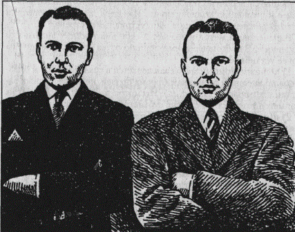
A special group among MBs are unusual types of twins: two-headed (as a rule, non-viable), caspophagus ("Siamese twins"). Born in 1811 in Siam (now Thailand), Siamese twins Chang and Eng lived 63 years. They were married to twins; Chang produced 10, and Eng produced 12 children. Chang died of bronchitis, and Eng died 2 hours later. They were tied together by a tissue web about 10 cm wide. Later it was found that this web was made up of liver tissue and connected two livers. Any surgical attempt to divide the brothers at that time would hardly have been successful. More complex bonds between the twins are also being disconnected.
Dizygotic twins (DB) develop if two eggs are fertilized at the same time, fertilized by two spermatozoa.
Naturally, dizygotic twins have different genotypes. They are similar to each other no more than brothers and sisters, since they have about 50% identical genes. The total frequency of birth of twins is about 1%; of these, about 1/3 are in monozygotic twins. It is known that the number of births of monozygotic twins is similar in different populations, while for dizygotic twins, this figure differs significantly. For example, in the US, dizygotic twins are born more often among blacks than whites. In Europe, the frequency of dizygotic twins is 8 per 1000 births. However, in some populations they are more. The lowest frequency of birth of twins, inherent in a greater degree of Mongoloid populations, is observed in Japan. It is noted that the frequency of congenital deformities in twins, as a rule, is higher than in single-born.
It is believed that multiple babies are genetically determined. However, this is true only for dizygotic twins. Factors affecting the frequency of birth of twins, are currently little studied. There are data showing that the probability of dysygotic twins birth increases with increasing age of the mother, as well as the serial number of birth. The effect of maternal age is probably explained by an increase in gonadotropin levels, leading to an increase in poliovulation. There is evidence of a decrease in the frequency of birth of twins in industrialized countries.
The twin method includes diagnosing the zygosity of twins. Currently the following methods are used to establish it:
1) The polysymptomatic method consists in comparing a pair of twins according to external signs (shape of eyebrows, nose, lips, auricles, hair color, eyes, etc.). Despite the obvious convenience, this method is to a certain extent subjective and may give errors;
2) the immunogenetic method is more complicated and is based on the analysis of blood groups, serum proteins, leukocyte antigens, sensitivity to phenylthiocarbamide, etc. If the twins do not differ by these signs, they are considered monozygous;
3) a reliable criterion for twin zygosity is the survival of pieces of skin. It has been established that in dizygotic twins such a transplant always ends in rejection, while in monozygous couples there is a high incidence of transplantation;
4) the method of dermatoglyphics consists in studying the papillary patterns of the fingers, palms and feet. These signs are strictly individual and do not change throughout a person’s life. It is not by chance that these indicators are used in forensic science and forensic medicine to identify the person and establish paternity. The similarity of dermatoglyphic indices in monozygotic twins is significantly higher than that of dizygotic.
The twin method also includes a comparison of groups of mono and dizygotic twins on the trait under study. If any sign is found in both twins of the same pair, then it is called concordant, if in one of them, then the pair of twins is called discordant (concordance is the degree of similarity, discordance is the degree of difference).
When comparing mono-and dizygotic twins, they determine the coefficient of pair concordance, indicating the proportion of twin pairs in which the studied trait manifested itself in both partners. The concordance factor is expressed in fractions of a unit or as a percentage and is determined
according to the formula: K = C / (C + D)
where C is the number of concord pairs
D - the number of discordant pairs.
Comparison of pair concordance in mono- and dizygotic twins gives an answer about the correlative role of heredity and the environment in the development of a particular trait or disease. At the same time, it is assumed that the degree of concordance is significantly higher in monozygous than in dizygotic twins, if hereditary factors play a dominant role in the development of the trait (see Table 1).
Table 1. Concordance of some signs of a person in identical (OB) and dual (DB) twins
| Signs of | Concordance,% | |
| ABOUT | Db | |
| Normal | ||
| Blood type | ||
| Eye color | 99,5 | |
| Hair color | ||
| Papillary patterns | ||
| Pathological | ||
| Clubfoot | ||
| "Rabbit lips" | ||
| Congenital dislocation of the hip | ||
| Paralytic poliomyelitis | ||
| Bronchial asthma | 4,8 | |
| Measles | ||
| Parotitis | ||
| Tuberculosis | ||
| Diphtheria | ||
| Epilepsy | ||
| Schizophrenia | ||
| Hypertension | ||
| Rheumatism | 20,3 | 6,1 |
If the value of the concordance coefficient is approximately close in monozygous and dizygotic twins, then they consider that the development of the trait is determined mainly by non-genetic factors, i.e. environmental conditions.
If both genetic and non-genetic factors are involved in the development of the trait, then monozygotic twins will have certain intra-pair differences. This will reduce the differences between mono - and dizygotic twins in the degree of concordance. In this case, it is considered that there is a genetic predisposition for the development of the trait. For a quantitative assessment of the role of heredity and the environment in the development of a trait, various formulas are used. Most often use the coefficient of heritability, which is calculated by the formulas:
Н = КМБ - КДБ: 100 - КДБ (in percentage);
Н = КМБ - КДБ: 1 - КДБ (in unit shares),
where H is the coefficient of heritability,
K - coefficient of pair concordance
in the group of monozygous or dizygotic twins.
Depending on the value of H, the influence of genetic and environmental factors on the development of the trait is judged. For example, if the value of H is close to zero, it is considered that the development of a trait is due only to environmental factors. When the value of H is from 1 to 0.7, hereditary factors are dominant in the development of a sign or disease; the average value of H from 0.4 to 0.7 indicates that the trait develops under the influence of environmental factors in the presence of a genetic predisposition.
Consider a few examples. As already noted, the blood groups in humans are completely determined by the genotype and do not change under the influence of the environment. Heritability coefficient is 100%. According to some morphological features (the shape of the nose, eyebrows, lips and ears, eye color, hair and skin), monozygotic twins are concordant in 97-100%, and dizygotic (depending on the trait) - in 70-20% of cases. The concordance of MB in terms of the incidence of schizophrenia is 70%, and in DB - 13%. Then H = (70 - 13): (100 - 13) - 0.65 or 65%. In this case, genetic factors prevail, but environmental conditions play a significant role.
Using the twin method revealed the value of the genotype and the environment in the pathogenesis of many infectious diseases. So, in case of measles and whooping cough disease, infectious factors are of primary importance, and in case of tuberculosis infection, the genotype has a significant effect. Studies conducted on twins will help answer questions such as the influence of hereditary and environmental factors on a person’s life expectancy, the development of giftedness, drug sensitivity, etc.
Currently, the twin method in human genetics is used in combination with other methods of genetic analysis.
Questions and tasks
1. What are the differences between monozygotic and dizygotic twins?
2. Give the main methods of diagnosis of mono-and di-gothic.
3. Apply the twin method to analyze alternative signs.
4. Give examples of the concordance of twins in certain infectious and polyfactorial diseases.
7.Biochemical methods. They allow you to identify changes in metabolism to clarify the diagnosis of the disease, heterozygous carrier state. For example, heterozygous carriers of the phenylketonuria recessive allele respond to the administration of phenylalanine by a stronger increase in the concentration of the amino acid in the plasma than normal homozygotes. This method is used in medical genetic counseling to determine the likelihood of having a child with a hereditary disease.
Diseases based on metabolic disorders constitute a significant part of hereditary pathology (phenylketonuria, galactosemia, alkaptonuria, etc.).
It is possible to assume that the patient has a hereditary defect of exchange by the following criteria:
1) mental retardation, isolated or in combination with the pathology of other organs;
2) violation of mental status;
3) impaired physical development;
4) seizures, muscle hypo or hypertension, gait disturbance and coordination of movements, jaundice, hypo or hyperpigmentation;
5) intolerance to certain foods and medicines, digestive disorders, etc.
8. Population-statistical method.
One of the important trends in modern genetics is population genetics. It studies the genetic structure of populations, their gene pool, the interaction of factors responsible for the constancy and change in the genetic structure of populations. A population in genetics is a collection of freely mating individuals of the same species occupying a specific arial and possessing a common gene pool in a number of generations. The gene pool is the entire set of genes found in individuals of a given population.
In medical genetics, the population-statistical method is used in the study of hereditary diseases of the population, the frequency of normal and pathological genes, genotypes and phenotypes in populations of various localities, countries and cities. In addition, this method studies the patterns of the spread of hereditary diseases in populations of different structure and the ability to predict their frequency in subsequent generations.
Population-statistical method is used to study:
The frequency of genes in a population, including the frequency of hereditary diseases;
Patterns of the mutation process;
The role of heredity and environment in the occurrence of diseases with hereditary predisposition;
The effects of hereditary and environmental factors in the creation of human phenotypic polymorphism in many ways, etc.
The use of the population-statistical method includes the correct selection of the population, the collection of material and the statistical analysis of the results obtained.
The method is based on the regularity established in 1908 by the English mathematician J. Hardy and the German doctor V. Weinberg for an ideal population. The pattern they discovered is called the Hardy-Weinberg law.
For an ideal population, the following features are characteristic: a large population size, free crossing (panmixia) of organisms, lack of selection and a mutation process, and absence of migrations into and out of the population. In an ideal population, the frequency ratio of dominant homozygotes (AA), heterozygotes (Aa), and recessive homozygotes (aa) remains constant from generation to generation if no evolutionary factors disturb this balance. This is the basic meaning of the Hardy-Weinberg law. When any of these equilibrium conditions change, the ratio of the number of genotypes in the population is disturbed. These conditions include related marriages, mutations, gene drift, selection, migration, and other factors. However, this does not reduce the importance of the Hardy-Weinberg law. It is the basis when considering genetic transformations occurring in natural and artificially created populations of plants, animals and humans.
The ratio of the number of different genotypes and phenotypes in a panmictic population is determined by the Newton binomial formula:
(p + q) 2 = p 2 + 2pq + q 2; (p + q) 2 = 1
where p is the frequency of the dominant allele A;
q is the frequency of the recessive allele a;
p 2 - the frequency of the AA genotype (homozygotes for the dominant allele);
q 2 - frequency of aa genotype (homozygotes for the recessive allele).
In accordance with the Hardy-Weinberg law, the frequency of dominant homozygotes (AA) is equal to the square of the probability of occurrence of the dominant allele, the frequency of heterozygotes (Aa) is the doubled product of the probability of occurrence of the dominant and recessive alleles. The frequency of occurrence of recessive homozygotes (aa) is equal to the square of the probability of a recessive allele.
Thus, the population-statistical method makes it possible to calculate in the human population the frequency of normal and pathological heterozygous genes, dominant and recessive homozygotes, as well as the frequency of normal and pathological phenotypes, i.e., to determine the genetic structure of the population.
An important factor affecting the frequency of alleles in small populations and in isolates are genetic-automatic processes, or gene drift. This phenomenon was described in the 30s. N. P. Dubinin and D. D. Romashev (USSR), S. Wright and R. Fisher (USA). It is expressed in random changes - the frequency of alleles that are not related to their selective value and the action of natural selection. As a result of gene drift, adaptive alleles can be eliminated from a population, and less adaptive and even pathological (for random reasons) can persist and reach high concentrations. As a result, a rapid and sharp increase in the frequencies of rare alleles can occur in a population.
Genetic-automatic processes most intensively occur with the uneven reproduction of individuals in the population. Fluctuations in population numbers are often observed in insects, rodents, and other animals in the form of so-called "waves of life." In some favorable years, their number greatly increases, and then drops sharply. The reasons may be the development of diseases, lack of food, lower temperatures, etc. As a result of a decline in the population size or in isolated populations, heterozygosity decreases and the genetic homogeneity of the population increases.
An example of the effect of gene drift in human populations is the “ancestor effect”. It is observed if the structure of the population is formed under the influence of alleles of a limited number of families. In such populations, a high frequency of an abnormal gene, preserved as a result of random gene drift, is often observed. It is possible that the result of gene drift is the different frequency of Rh-negative people in Europe (14%) and in Japan (1%), the uneven spread of hereditary diseases in different populations of the globe. For example, in some populations in Sweden, the gene of juvenile amarotic idiocy is widely distributed, in South Africa - the gene of porphyria, in Switzerland - the gene of hereditary deafness, etc.
Closely related marriages (inbreeding) significantly affect the genotypic composition of the population. Such marriages are most often made between a niece and an uncle, cousin brother and sister. Closely related marriages are prohibited in many countries. This is due to the high probability of having children with hereditary pathology. Relatives, having a common origin, can be carriers of the same recessive pathological gene, and if two healthy heterozygotes are married, the probability of having a sick child becomes high.
New genes can enter the population as a result of migration (gene flow), when individuals from one population move to another and interbreed with representatives of this population. Real populations are rarely completely isolated. There is always some movement of individuals from one population to another. It can be not only active, but also passive (transfer of seeds by birds). Sometimes a person deliberately mixes populations. For example, in Siberia, in order to improve local sables, Barguzin sables are produced in their populations with very dark fur coloration, more valued in the fur industry. This leads to a change in the frequency of alleles in the main population and among "immigrants". In local populations, the frequency of alleles can vary if the old-timers and aliens have different initial frequencies of alleles. Similar processes occur in human populations.
In the US, offspring from mixed marriages between whites and blacks belongs to the Negro population. According to F. Ayala and J. Kiger (1988), the frequency of the allele controlling the Rh factor in the white population is 0.028. In African tribes, from which the modern Negro population originates, the frequency of this allele is equal to 0.630. The ancestors of the modern blacks of the United States were exported from Africa 300 years ago (about 10 generations). The frequency of the allele in the modern Negro population of America is 0.446. Thus, the flow of genes from the white to the Negro population went at a rate of 3.6% per generation. As a result, after 10 generations, the share of genes of African ancestors is now 0.694 of the total number of genes of the modern Negro population of the USA. About 30% of the genes of American negros inherited from the white population. Obviously, the flow of genes between the white and Negro population was significant.
Finally, it should be briefly examined how the mutational process and selection influence the genetic structure of populations. Mutations as a factor of evolution provide an influx of new alleles in the population. By genotype change, mutations are divided into gene (or point), intrachromosomal and interchromosomal, genomic (change in the number of chromosomes.). Gene mutations can be direct (A) and reverse (A). The frequency of direct mutations is much higher than the reverse. The same genes can mutate many times. In addition, the same gene can change into several allelic states, forming a series of multiple alleles (A a1, a2, a3, an). The study of the frequency of mutations that cause such serious diseases in humans as hemophilia, retinoblastoma, xeroderma pigment, etc., suggests that the frequency of occurrence of pathological mutations of a single gene is about 1-2 per 100 thousand gametes per generation. Given the total number of genes in humans (about 100 thousand), the total mutability is a considerable amount. The frequency of mutations can significantly increase with the action on the body of certain physical and chemical factors (mutagens). Chemical mutagens are found among industrial poisons, insecticides, herbicides, food additives and drugs. Most carcinogens also have mutagenic effects. In addition, many biological factors, such as viruses and live vaccines, as well as histamine and steroid hormones produced in humans, can induce mutations. Strong mutagens are various types of radiation (X-rays and γ-rays, β-particles, neutrons, etc.) that can cause gene and chromosomal mutations in humans, as evidenced by the consequences of the Chernobyl nuclear power plant accident.
The factors that violate the constancy of the genetic structure of the population include natural selection, which causes a directional change in the gene pool by eliminating from a population of less adapted individuals or reducing their fertility. Consider the effect of selection on the example of the dominant pathology - achondroplasia (dwarfism). This disease is well studied in Danish populations. Patients have reduced vitality and die in childhood, i.e., are eliminated by natural selection from the population. Surviving dwarfs are less likely to marry and have few children. The analysis shows that about 20% of the achondroplasia genes are not transmitted from parents to children, and 80% of these genes are eliminated from the population. From these data it follows that achondroplasia does not have a significant effect on the structure of the population.
Most mutant genotypes have low selective value and are subject to selection. According to V. Makkyusika (1968), about 15% of fetuses die before birth, 3% of children die before reaching puberty, 20% die before marriage, in 10% of cases the marriage remains fruitless.
However, not every mutant gene decreases selective
1. Genealogical method. 2. Twin method. 3. Method of dermatoglyphics. 4. Cytogenetic method. 5. Method of hybridization of somatic cells. 6. Entogenetic method. 7. Population-statistical method. 8. Method of modeling. 9. Immunological method. .Biochemical method.
Genealogical method: or the study of pedigrees provides for tracing the trait in the family or genus, indicating the type of kinship between the members of the pedigree. In medical genetics, this method is commonly referred to as clinical and genealogical, since it is about observing pathological signs using techniques of clinical examination. The genealogical method refers to the most universal methods in human genetics. It is widely used in solving theoretical and practical problems. The essence of the genealogical method comes down to clarifying kinship and tracing the trait among near and far direct and indirect relatives. Technically, it consists of two stages: the compilation of pedigrees and genealogical analysis.
Twin study method: The study of twins - one of the main methods of human genetics. There are identical twins, arising from a single egg fertilized by a single sperm cell. They arise because of the separation of zygotes into two genetically identical and always same-sex embryos. Multiple egg twins develop from different eggs fertilized by different sperm cells. Genetically, they differ as brothers and sisters of the same parents.
Using the twin method can be studied:
1) The role of heredity and environment in the formation of the physiological and pathological features of the organism. In particular, the study of the hereditary transmission of certain diseases by people. The study of expressiveness and penetrance of genes that cause hereditary diseases.
2) Specific factors that enhance or weaken the influence of the external environment.
3) Correlation of features and functions.
Dermatoglyphics method: uhthis is the science that studies the hereditary conditionality of the patterns that form the skin lines on the fingertips, the palms and the soles of the person.
It turned out that every nation, every race, every person has their own patterns, and they are strictly individual on the palms. F. Galton first drew attention to this, who suggested that the British criminal police identify the criminals by fingerprints. Dermatoglyphic studies are important in forensic science, in determining the zygosity of twins, in the diagnosis of a number of hereditary diseases, as well as in some cases of controversial paternity.
Cytogenetic method: This method allows using a microscope to investigate the structure of the cell - chromosome. Using the method of microscopy, the karyotype of the human body (chromosome set of body cells) was studied. It has been established that many diseases and developmental defects are associated with a violation of the number of chromosomes and their structure. This method also makes it possible to study the effect of mutagens on the composition and structure of chromosomes.
Somatic cell hybridization: Hybrid cells have certain properties that allow you to determine the localization of a gene or gene linkage. The loss of human chromosomes from some types of hybrid cells allows to obtain clones with the absence of a specific chromosome. The most commonly used hybrids of human somatic cells are mouse.
Ontogenetic method: Allows you to study the patterns of manifestation of any sign or disease in the process of individual development. There are several periods of human development. Antenatal (development before birth) and postnatal.
Population-statistical research method: It is a method of mathematical calculation of various genes and corresponding traits in certain populations. The theoretical basis of this method is the Hardy-Weinberg law.
Simulation method The law of N.I. Vavilov’s homologous series (genetically close species and genera have similar series of hereditary variability) allows extrapolating experimental data to humans with certain limitations.
Immunological research method : This method is based on the study of the antigenic composition of cells and body fluids of the human body - blood, saliva, gastric juice, etc. The most common antigens are blood cells: red blood cells, white blood cells, platelets, as well as blood proteins. Different types of erythrocyte antigens form blood group systems.
Biochemical method: Allows, on the one hand, to study the amount of DNA in human cells in health and disease, on the other - to determine hereditary metabolic defects. prp phenylketanuria
Examination ticket number 32
1. Factors of microevolution. Elementary evolutionary factors:
- Mutation process (Constant mutational variability and combinations at crossings give new combinations of genes in the gene pool, which inevitably causes hereditary changes in the population. The mutation process constantly increases the genetic heterogeneity of a population, due to the persistence of recessive mutations in heterozygotes. But the mutation process itself without the participation of other factors of evolution can not direct the change in the natural population. He is only a supplier of elementary evolutionary material.
-Population waves. Gene drift ( Population waves are periodic fluctuations in the number of individuals in a population. The reasons for these fluctuations can be various abiotic and biotic environmental factors. With a sharp reduction in population size (seasonal fluctuations, reduction in resources), there may be rare genotypes among the few surviving individuals. If in the future the number is restored by these individuals, then this will lead to an accidental change in the frequency of the genes in the gene pool. Thus, population waves are the supplier of evolutionary material. If the population is small in size, as a result of random events, some individuals, regardless of their genetic constitution, can leave or not leave offspring, as a result of which the frequencies of some genes can change dramatically over a single generation. A random change in the frequency of genes in the gene pool of a population is called gene drift.
-Insulation (due to the emergence of a variety of factors that impede the free crossing. Reproduction occurs mainly within the isolate, the exchange of genetic information with other groups ceases. This contributes to the consolidation of the initial stage of changing the gene pool of a separate group, its formation as an independent genetic system. There are spatial and biological isolation.
Spatial isolation is associated with geographically (water barriers, mountain ranges, places unsuitable for life) and environmental (resettlement in different ecological niches) factors of population separation. The value of spatial isolation depends on the size of the individual activity of individuals of the species.
Biological isolation may include behavioral patterns, changes in the structure and physiological activity, the timing of reproduction, and a number of other factors that interfere with mating. After fertilization, violations of chromosome conjugation and a number of other changes are possible, leading to the development of fully or partially sterile hybrids, as well as hybrids with reduced viability. The evolutionary significance of different forms of isolation is that it reinforces and enhances genetic differences between populations.
Changes in gene frequencies caused by the evolution factors discussed above are random, non-directional, and even their combined action does not lead to the sustainable implementation of the directional process of evolution. The guiding factor is natural selection. .-Natural selection (Darwin’s natural selection principle is fundamental to evolutionary theory. Natural selection is the directional driving force behind the evolution of the organic world. Nowadays, natural selection has been replenished with new facts that have expanded and deepened. Natural selection should be understood as selective survival and the possibility of leaving separate descendants individuals. The biological value of an individual that has given offspring is determined by the contribution of its genotype to the gene pool of a population. Its objects are the phenotypes of individual individuals.The organism's phenotype is formed on the basis of the implementation of the information of the genotype in certain environmental conditions.
The use of this method is possible in the case when direct relatives are known - the ancestors of the owner of the hereditary trait ( proband) on the maternal and paternal lines in a series of generations or descendants of the proband also in several generations. In the compilation of genealogies in genetics uses a specific notation. After compiling the pedigree, it is analyzed in order to establish the nature of the inheritance of the trait under study.
Conventions used in compiling pedigrees:
1 - the man; 2 - a woman; 3 - gender not clarified; 4 - the owner of the studied trait; 5 - heterozygous carrier of the recessive gene under study; 6 - marriage; 7 - marriage of a man with two women; 8 - related marriage; 9 - parents, children and the order of their birth; 10 - dizygotic twins; 11 - monozygotic twins.
Due to the genealogical method, the types of inheritance of many traits in humans were determined. Thus, in an autosomal dominant type, polydactyly (increased number of fingers), the ability to roll the tongue into a tube, brachydactyly (short flank due to the absence of two phalanges on the fingers), freckles, early baldness, accrete fingers, cleft lip, cleft palate, cataract eyes, are inherited the fragility of bones and many others. Albinism, red hair, susceptibility to poliomyelitis, diabetes mellitus, congenital deafness and other signs are inherited as autosomal recessive.
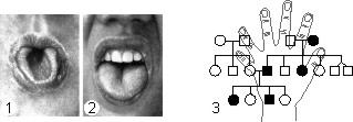
The dominant trait is the ability to roll the tongue into a tube (1) and its recessive allele - the absence of this ability (2).
3 - pedigree on polydactyly (autosomal dominant inheritance).
A number of signs are inherited sexually: X-linked inheritance - hemophilia, color blindness; Y -linked - hypertrichosis of the edge of the auricle, webbed toes. There are a number of genes localized in homologous regions of the X - and Y -chromosomes, for example, general color blindness.
The use of the genealogical method has shown that with a related marriage, compared with an unrelated, the probability of the appearance of deformities, stillbirths, and early mortality in offspring significantly increases. In related marriages, recessive genes are more likely to become homozygous, with the result that certain anomalies develop. An example of this is the inheritance of hemophilia in the royal houses of Europe.
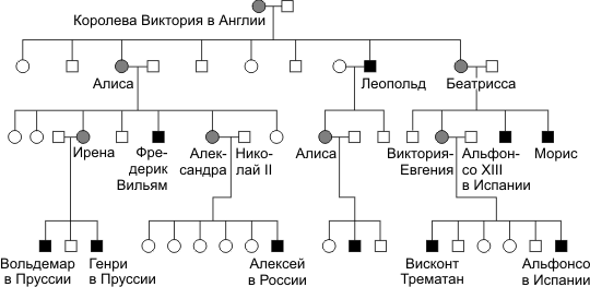
- hemophilic; - a female carrier.
This method is used in human genetics to determine the degree of hereditary dependence of the studied traits. Gemini can be identical (formed in the early stages of crushing zygotes, when from two or less from a larger number of blastomeres develop high-grade organisms). Identical twins are genetically identical. When more or more eggs are fertilized by various spermatozoa and then fertilized with different spermatozoa, runny twins develop. Fraternal twins are similar to each other no more than brothers and sisters born at different times. The frequency of occurrence of twins in humans is about 1% (1/3 of the identical, 2/3 of the fraternal); the vast majority of twins are twins.
Since the hereditary material of identical twins is the same, the differences that arise from them depend on the influence of the environment on gene expression. Comparison of the frequency of similarity in a number of signs of pairs of single and fraternal twins allows us to estimate the value of hereditary and environmental factors in the development of the human phenotype.
Twins are called simultaneously born children. They are monozygous (identical) and dizygotic (fraternal).
Monozygous twins develop from one zygote (1), which at the crushing stage was divided into two (or more) parts. Therefore, these twins are genetically identical and always of the same sex. Monozygous twins are characterized by a high degree of similarity ( by concordance) in many ways.
Dizygotic twins develop from two or more ovulified and fertilized eggs with different sperm (2). Therefore, they have different genotypes and can be both single and different sexes. Unlike monozygous, dizygotic twins are characterized by discordance - dissimilarity in many ways. Data on the concordance of the twins on some grounds are given in the table.
| Signs of | Concordance,% | |
|---|---|---|
| Monozygous twins | Dizygotic twins | |
| Normal | ||
| Blood type (AB0) | 100 | 46 |
| Eye color | 99,5 | 28 |
| Hair color | 97 | 23 |
| Pathological | ||
| Clubfoot | 32 | 3 |
| “Rabbit's lip” | 33 | 5 |
| Bronchial asthma | 19 | 4,8 |
| Measles | 98 | 94 |
| Tuberculosis | 37 | 15 |
| Epilepsy | 67 | 3 |
| Schizophrenia | 70 | 13 |
As can be seen from the table, the degree of concordance of monozygotic twins in all the above characteristics is significantly higher than that of dysygotic ones, but it is not absolute. As a rule, the discordance of monozygotic twins arises as a result of violations of the intrauterine development of one of them or under the influence of the external environment, if it was different.
Thanks to the twin method, hereditary predisposition of a person to a number of diseases was found out: schizophrenia, epilepsy, diabetes and others.
Observations of monozygotic twins provide material for clarifying the role of heredity and the environment in the development of traits. Moreover, the external environment means not only physical environmental factors, but also social conditions.
Methods of population genetics are widely used in human studies. Intra-familial morbidity analysis is inseparable from the study of hereditary pathology, both in individual countries and in relatively isolated groups of the population. The study of the frequency of genes and genotypes in populations is the subject of population genetic research. This gives information about the degree of heterozygosity and polymorphism of human populations, reveals differences in allele frequencies between different populations.
Considered to be Hardy's law? Weinberg suggests that inheritance as such does not change the frequency of alleles in the population. This law is quite suitable for the analysis of large populations where there is a free crossing. The sum of the frequencies of alleles of one gene, according to the formula of Hardy? Weinberg p + q = 1, in the gene pool of a population is a constant value. The sum of the frequencies of the genotypes of the alleles of a given p2 + 2pq + q2 = 1 gene is also a constant value. With full dominance, by setting the number of recessive homozygotes (q2? Number of homozygous "individuals by the recessive gene with aa genotype) in this given population, it suffices to extract the square root of the obtained value, and we find the frequency of the recessive allele A. The frequency of the dominant allele A will be p = 1 - q. By calculating the frequencies of alleles a and A, you can determine the frequencies of the corresponding genotypes in the population (p2 = AA; 2pq = Aa). For example, according to a number of scientists, the frequency of albinism (inherited as an autosomal recessive trait) is 1 : 20 000 ( q2). Therefore, the frequency of allele a in the gene pool will be q2 = l / 20000 = / l4l and then the frequency of allele A will be
p = 1-q. p = 1. p = 1-1 / 141 = 140/141.
In this case, the frequency of heterozygous carriers of the albinism gene (2pq) will be 2 (140/141) x (1/141) = 1/70, or 1.4%
Statistical analysis of the distribution of individual hereditary traits (genes) in populations of people in different countries allows us to determine the adaptive value of specific genotypes. Once having arisen, mutations can be transmitted to offspring for many generations. This leads to polymorphism (genetic heterogeneity) of human populations. Among the population of the Earth it is almost impossible (with the exception of identical twins) to find genetically identical people. A significant number of recessive alleles (genetic load) causing the development of various hereditary diseases are in the heterozygous state in the populations. The frequency of their occurrence depends on the concentration of the recessive gene in the population and increases significantly when entering into closely related marriages.
In 1892 F.Galton as one of the methods for the study of man was proposed a method of studying the scallop skin patterns of the fingers and palms, as well as flexion palmar furrows. He found that these patterns are an individual characteristic of a person and do not change throughout life.At present, hereditary conditionality of skin patterns has been established, although the nature of inheritance has not been finally clarified. Probably, the trait is inherited according to a polygenic type. Dermatoglyphic studies are important in identifying twins. The study of people with chromosomal diseases revealed specific changes in them, not only of the patterns of fingers and palms, but also of the nature of the main bending grooves on the skin of the palms. Dermatoglyphic changes in gene diseases are less studied. Basically, these methods of human genetics are used to establish paternity.
Examination of the prints of the skin pattern of the palms and feet. With the existing individual differences in the fingerprints due to the peculiarities of the development of the individual, several main classes of them are distinguished.Peculiar changes of fingerprints and palm patterns were observed in a number of hereditary degenerative diseases of the nervous system.Typical of Down's disease is a monkey (four-fingered) fold, representing a line running across the palm in the transverse direction.Currently, the method is mainly used in forensic medicinee.
Hereditary diseases that are caused by gene mutations that change the structure or rate of protein synthesis, are usually accompanied by a violation of carbohydrate, protein, lipid and other types of metabolism. Hereditary metabolic defects can be diagnosed by determining the structure of the altered protein or its quantity, identifying defective enzymes or detecting the intermediate metabolic products in the extracellular body fluids (blood, urine, sweat, etc.). For example, the analysis of amino acid sequences of mutationally altered protein chains of hemoglobin revealed several hereditary defects underlying a number of diseases? hemoglobinosis. Thus, in sickle cell anemia in humans, abnormal hemoglobin due to mutation differs from the normal one by replacing only one amino acid (glutamic acid with valine).
In the practice of health care, in addition to identifying homozygous carriers of mutant genes, there are methods for identifying heterozygous carriers of certain recessive genes, which is especially important for medico-genetic counseling. Thus, phenotypically normal heterozygotes for phenylketonuria (a recessive mutant gene; amino acid metabolism of phenylalanine is disturbed in homozygotes, which leads to mental retardation) after ingestion of phenylalanine, its elevated blood levels are detected. In hemophilia, heterozygous carriage of the mutant gene can be established by determining the activity of the enzyme that has been altered as a result of the mutation.
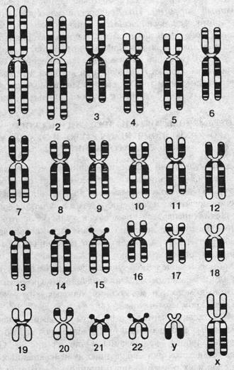
The cytogenetic method is used to study the normal human karyotype, as well as in the diagnosis of hereditary diseases associated with genomic and chromosomal mutations. In addition, this method is used in the study of the mutagenic action of various chemicals, pesticides, insecticides, drugs, etc.
During the period of cell division into metaphase stages, chromosomes have a clearer structure and are available for study. The diploid set of a person consists of 46 chromosomes: 22 pairs of autosomes and one pair of sex chromosomes (XX? In women, XY? In men). Usually, human peripheral blood leukocytes are examined, which are placed in a special nutrient medium, where they divide. Then prepare the preparations and analyze the number and structure of the chromosomes. The development of special coloring methods significantly simplified the recognition of all human chromosomes, and together with the genealogical method and the methods of cell and genetic engineering, made it possible to correlate genes with specific chromosome regions. The integrated application of these methods underlies the mapping of human chromosomes. Cytological monitoring is necessary for the diagnosis of chromosomal diseases associated with ansuploidy and chromosomal mutations. The most common Down syndrome (trisomy 21 st chromosome), Klinefelter syndrome (47 XXY), Shershevsky syndrome? Turner (45 XO) and others. Loss of a portion of one of the homologous chromosomes of the 21st pair leads to blood disease? chronic myeloid leukemia.
Cytological studies of the interphase nuclei of somatic cells can reveal the so-called Barry body, or sex chromatin. It turned out that sex chromatin is normal in women and absent in men. It is the result of heterochromatization of one of the two X chromosomes in women. Knowing this feature, you can identify gender and identify an abnormal number of X chromosomes.
Detection of many hereditary diseases is possible even before the birth of a child. The method of prenatal diagnosis is to obtain amniotic fluid, where the cells of the fetus are located, and in the subsequent biochemical and cytological determination of possible hereditary anomalies. This allows you to make a diagnosis in early pregnancy and make a decision about her continuation or termination.
With the help of these methods, the heredity and variability of somatic cells are studied, which compensates for the impossibility of applying a hybridological analysis to a person. These methods, based on the reproduction of these cells in artificial conditions, analyze the genetic processes in individual cells of the body, and thanks to the usefulness of the genetic material, use them to study the genetic patterns of the whole organism.
In genetic studies of humans use the following techniques:
Hybrid cells containing 2 complete genomes usually divide chromosomes, preferably one of the species, upon division. Thus, cells with the desired set of chromosomes can be obtained, which makes it possible to study the linkage of genes and their localization in certain chromosomes.
Thanks to the methods of somatic cell genetics, it is possible to study the mechanisms of the primary action and interaction of genes, the regulation of gene activity. The development of these methods determined the possibility of accurate diagnosis of hereditary diseases in the prenatal period.
Somatic cell genetics studies the heredity and variability of somatic cells, i.e. cells of the body, not genital. Somatic cells have the entire set of genetic information, they can study the genetic characteristics of the whole organism.
Human somatic cells are obtained for genetic studies from the material biopsies (intravital excision of tissues or organs) when a small piece of tissue is taken for examination. As a rule, this is done during operations, when it is necessary to establish whether a given formation, for example, a tumor, has a malignant or benign nature.
Currently used the following methods of somatic cell genetics : simple cultivation, hybridization, cloning and selection. Simple cultivation - this is the multiplication of cells on nutrient media to get them in sufficient quantity for cytogenetic, biochemical, immunological and other methods.
With somatic cell hybridization You can cross cells derived from different people, as well as human cells with cells of mice, rats, guinea pigs, monkeys and other animals. Such studies make it possible to establish linkage groups, and using chromosomal rearrangements to identify the sequence of the location of genes and build genetic maps of human chromosomes.
Cloning - this is getting offspring of one cell (clone). All cells as a result of cloning will be the same genotype.
Selection - this is the selection of cells with predetermined properties. Then the cultivation and reproduction of these cells on special nutrient media. For example, you can use a nutrient medium without lactose, but with the addition of other sugars, and from a large number of cells placed in it, there may be several that can live in the absence of lactose. Then a clone is obtained from such cells.
Studying human diseases in animals that can suffer from these diseases. It is based on the Vavilov law on homologous series of hereditary variability, for example, hemophilia linked to sex, can be studied in dogs, epilepsy in rabbits, diabetes, muscular dystrophy in rats, and lip and palate cleft in mice
Models in biology are used to model biological structures, functions, and processes at different levels of the organization of life: molecular, subcellular, cellular, organ-systemic, organismic, and population-biocenotic. It is also possible to model various biological phenomena, as well as the living conditions of individual individuals, populations and ecosystems.
In biology, three types of models are mainly used: biological, physico-chemical, and mathematical (logical-mathematical). Biological models reproduce certain conditions or diseases found in humans or animals in laboratory animals. This makes it possible to study experimentally the mechanisms of the occurrence of a given condition or disease, its course and outcome, to influence its course. Examples of such models are artificially induced genetic disorders, infectious processes, intoxication, reproduction of the hypertensive and hypoxic states, malignant neoplasms, hyperfunction or hypofunction of certain organs, as well as neuroses and emotional states. To create a biological model, various methods of influence on the genetic apparatus, infection with microbes, the introduction of toxins, the removal of individual organs or the introduction of their metabolic products (for example, hormones), various effects on the central and peripheral nervous systems, exclusion of certain substances from food, in artificially created habitat and many other ways. Biological models are widely used in genetics, physiology, and pharmacology.
The method of modeling in biology is a means to establish all the deeper and complex relationships between biological theory and experience. In the last century, an experimental method in biology began to run into certain boundaries, and it turned out that a whole series of studies was impossible without modeling. If we look at some examples of limitations of the scope of the experiment, they will be mainly as follows: (19 с15)
Experiments can be carried out only on the currently existing facilities (the impossibility of extending the experiment to the area of the past);
Intervention in biological systems is sometimes of such a nature that it is impossible to establish the reasons for the changes that have appeared (due to intervention or for other reasons);
Some theoretically possible experiments are not feasible due to the low level of development of experimental technology;
A large group of experiments related to human experimentation should be rejected for moral and ethical reasons.
But modeling is widely used in the field of biology, not only because it can replace the experiment. It has great independent value, which is expressed, according to a number of authors (19, 20,21), in a number of advantages:
1. Using the simulation method on one set of data, you can develop a number of different models, interpret the phenomenon under study in different ways, and choose the most fruitful of them for theoretical interpretation;
2. In the process of building a model, you can make various additions to the hypothesis under study and get its simplification;
3. In the case of complex mathematical models, computers can be used;
4. the possibility of conducting model experiments (the synthesis of amino acids according to Miller) (19 с152).
All this clearly shows that modeling performs independent functions in biology and is becoming an increasingly necessary step in the process of creating a theory. However, modeling retains its heuristic value only when the limits of application of any model are taken into account.
The immunogenetic method includes serological methods, immunoelectrophoresis, etc., which are used to study blood groups, proteins and serum enzymes of tissues. It can be used to establish immunological incompatibility, identify immunodeficiency, mosaicism of twins, etc.
An important part of medical genetics is immunogenetics, in particular, genetics of blood groups. Currently there are many major systems of blood groups. Of these, the AB0 and Rhesus systems are the most studied. For example, consider the Rhesus blood system. In the human genotype there is a dominant gene that determines the formation in the body of a special protein calledrh factor. A person who is homozygous (Rh + Rh +) or heterozygous (Rh + rh–) for this trait is Rh-positive, i.e. has this protein in the blood. And in the case of homozygosity for the recessive allele (rh– rh–), the Rh factor in the blood is absent. And if he enters the blood of such a person (during blood transfusion or pregnancy), a protective reaction develops in his body - like on any foreign protein, and specific antibodies are formed. Statistics show that among Europeans about 85% of people are Rh-positive and only 15% are Rh-negative.
In marriages of Rh-negative women (rh– rh– genotype) with Rh-positive homozygous men (Rh + Rh + genotype) due to the dominance of Rh-positive fetus (Rh + rh– genotype) is Rh-positive and releases Rh factor against which in the body mothers produce antibodies, which in turn destroy the fetal hematopoietic system. As a result, both the mother’s body and the fetus’s body suffer during pregnancy.
A similar situation may also occur in a Rh-negative woman’s marriage with a Rh-positive man heterozygous for this trait (genotype Rh + rh–). However, in this case, the probability of Rh conflict is halved, because the fetus may be Rh-negative (rh – rh– genotype), and then there is no conflict with the mother’s body.
In addition to the likelihood of the Rhesus conflict itself, which is determined solely by the genotypes of the parents, the degree of severity of the developing reaction is also important. In some cases, Rhesus-conflict occurs almost imperceptibly, in others it can cause the death of a child. Usually more severe consequences are observed during the second and subsequent pregnancies.
Currently, for the treatment of jaundice of newborns, caused by Rh-conflict, after birth, a full blood transfusion is performed to the child.
| Related articles: | |
|
Annual tax on cars
That each owner of the vehicle, ... Methods for the study of heredity in humans
1. Genealogical Genealogical method is to analyze ... Cauliflower puree soup with vegetables and cream
Today on our site is a recipe for cream of cauliflower soup with cream and ... | |


